Dorothy Keane, Clinical Lead at the Society and College of Radiographers, gives a brief overview of updates that have been made to our Clinical Imaging elearning programme.
The online training is free to access for healthcare staff and is the ideal resource to support all imaging staff.
Adult Pathology Sessions
“As radiographers, you are constantly looking at images of patients who have been referred from the emergency department, ward, outpatients, or a GP. Having the knowledge to recognise and identify bony changes which may represent a pathology will enable you to ‘flag’ the images to allow a faster report and quicker referral to a specialist”. Dorothy Keane, Clinical Lead
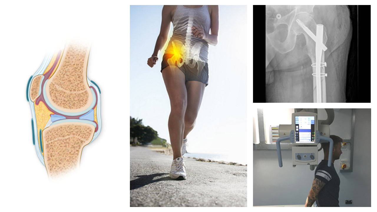
A new module has been developed to complement our Clinical Imaging programme. We have created 12 new sessions which give a general outline of a wide range of conditions and diseases and the related pathophysiological changes encountered on radiographs. These pathologies are commonly seen on radiographs and a knowledge of how bone and soft tissue changes manifest on radiographs will be discussed using images and diagrams. There is an opportunity to assess learning throughout each session which consist of 4 introductory sessions and 8 which focus on specific anatomical regions and discuss specific pathologies related to those regions.
Clinical Imaging – Orthopaedics
Have you looked at the 2 orthopaedic modules in elfh’s Clinical Imaging programme?
Our Orthopaedic Imaging modules explore follow-up images post orthopaedic surgery. The introductory session explains the post-operative plan for patients who have undergone orthopaedic procedures and why imaging is essential to assess the interventions.
Further sessions cover procedures involving the hip, knee, shoulder, elbow, ankle and foot, hand and wrist, long bones, vertebral column (spine) and pelvis in both emergency trauma and elective surgery. Each procedure is described with accompanying photographs of the prosthetics and instrumentation. The rationale for carrying out the procedure is discussed. Images are used to demonstrate post-procedure appearances and describe post-operative complications such as loosening of metalwork and infection.
The 2nd module, Orthopaedic Intervention, introduces the operating theatre outlining the environment, equipment, sterile procedures, infection control and staff roles. Radiographers often rotate into theatre and may have limited experience of certain procedures – this can, and often does, create an atmosphere of tension within the operating theatre for both the radiographer and the orthopaedic surgeons. These sessions have been designed to help prepare radiographers for theatre work. It provides detailed advice on the position and movements of the image intendifier for a range of orthopaedic procedure involving the upper and lower limbs and the vertebral column.
Accessing the training
To find out more and access the training, visit the Clinical Imaging programme page.


Comments are closed.