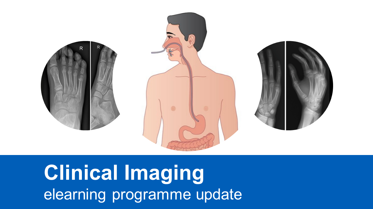
Dorothy Keane, Clinical Lead at the Society of Radiographers, gives a brief overview of several updates that have been made to our Clinical Imaging elearning programme. The online training is free to access for healthcare staff and is the ideal resource to support all imaging staff.
To find out more and access the training, visit the Clinical Imaging programme page.
Nasogastric Tube Placement
In 2017, Clinical Imaging developed 2 sessions which covered the main principles of correctly identifying a nasogastric (NG) tube position on a chest radiograph. The sessions were authored by Natasha Hayes who is a radiographer at Homerton University Hospital NHS Foundation Trust.
In April 2023, Natasha kindly reviewed and updated the sessions to include recent data and guidelines from a range of organisations including the National Patient Safety Agency (NPSA), Healthcare Safety Investigation Branch (HSIB) and the NHS Never Events data.
Natasha has also increased the number of chest radiographs in the Self Evaluation section to further test your knowledge on identifying correct placement of NG tubes. A big thank you to everyone involved.
Paediatric Imaging
The paediatric sessions in Clinical Imaging have recently been reviewed and updated. The sessions cover the appendicular and axial skeleton and focus on trauma, however common pathologies and normal variants are also included. The Suspected Physical Abuse (SPA) module has also been updated; it comprises of 12 sessions discussing the appearance of SPA on radiographs. The 1st session covers the knowledge and skills needed to recognise SPA. This is followed by 11 interactive case studies to test your understanding where you will decide whether the case is SPA or not.
New Paediatric Self Evaluation module
Self evaluation within Clinical Imaging has always been a major component of our programme. I decided to increase the number of sessions linked to paediatrics and to change the format. We now have 6 sessions covering the axial and appendicular skeleton as it appears on conventional radiographs. Each session is a combination of diagrams and images to label, hot spot questions, images to interpret and many other interactive questions covering anatomy, mechanisms of injury, radiographic technique as well as the interpretation of trauma, pathology and normal variants.
I feel that this update will enable you to complete a more thorough assessment of your skills in paediatric image interpretation.


Comments are closed.